ProCon X-Ray
- X-ray microscopes
Our CT Microscopes offer the perfect solution for situations where you need more than just high resolution.
These systems are specially developed for the highest resolution available on the market.
- CT-ALPHA Nanotube
- CT-NANO
CT-ALPHA Nanotube
CT-NANO
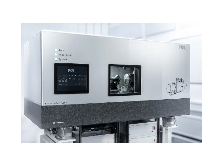
X-ray microscope CT-ALPHA Nanotube
CT-system for resolutions down to 150 nm.
The CT-ALPHA nanotube provides a unique solution for high-resolution measurements far ahead of established industrial micro CT scanners by applying the latest developments from X-ray research.
Features and parameters of the device:
- Min. voxel sampling 50 nm;
- Spatial resolution of 150 nm;
- Photon counting detector;
- Variable field of view;
- Automatic alignment;
- State-of-the-art reconstruction;
- CT-system for resolutions down to 150 nm
CT-ALPHA Nanotube system combines the latest state-of-the-art components for the highest resolution. Our experience in both hardware and software design enables us to perfectly adapt systems to your individual needs. Besides using only high-precision components, we also develop state-of-the-art reconstruction algorithms.
One main component of the system is Excillum’s high performance X ray source NanoTube N3, an up to 160 kV X-ray tube with latest tungsten-diamond transmission target technology, automatic beam focusing and astigmatism correction, ensuring that the smallest possible, truly round X-ray spot is achieved.
The second main component is the photon counting X ray detector from DECTRIS This detector enables an optimum signal-to-noise ratio due to zero readout noise and zero dark current.
Two separate manipulation systems allow a very precise detector and sample positioning. A high-precision air bearing rotary stage delivers a well-defined sample rotation, mounted on top of a linear position system with 3 degrees of freedom (DOFs). A separate manipulation (2 DOFs) system on top of the rotation stage is installed for automatically adjusting the sample position in the center of the rotation.
Specifications ProCon X-Ray CT-ALPHA Nanotube
|
Min. Min. voxel sampling |
50 nm |
|
Max. geometric magnification |
Up to 1,500 x for CT |
|
Detector |
Zero dark current |
|
Active area |
2,000 x 500 pikseli; 75 µm rpixel size |
|
High efficiency even on low energies |
For low contrast samples |
|
Variable field of view |
100 µm – 10 mm |
|
Spatial resolution |
down to150 nm |
|
Max. voltage |
up to 160 kV |
|
Axis system |
12-axes manipulator |
|
Axis resolution |
< 100 nm |
|
Automatic alignment |
Easy-to-use workflow |
|
State-of-the-art reconstruction |
Implemented |
|
Compact design |
Small footprint of 2.0 m x 1.0 m |
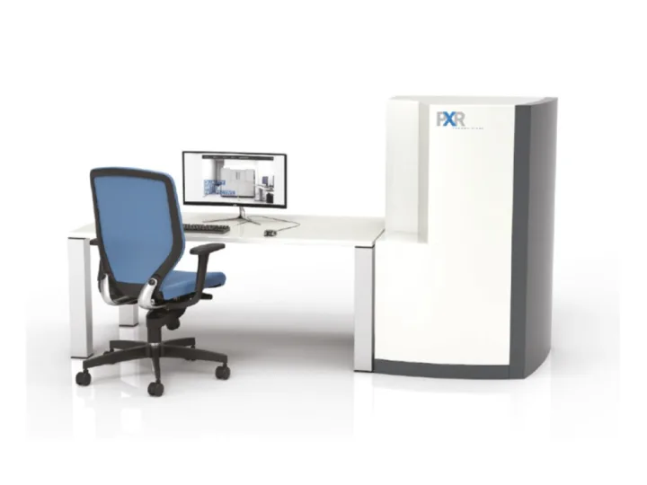
X-ray microscope CT-NANO
Explore new possibilities for visualization with 3D X-ray imaging for materials research, life sciences, natural resources and industrial applications.
- X-ray Computed Tomography (nanoCT);
- Scanning Electron Microscope (SEM);
- Digital Radiography (DR);
- Energy-Dispersive X ray spectroscopy (EDX);
- 3D volume CT;
- Non-destructive testing (NDT) – 2D and 3D;
- Quality control independent of material;
- Defect recognition (voids, cracks, …);
- Radiation safety better than 1 µSv/h.
CT-NANO PROCON X-Ray has the best resolution available on the market at this energy level, which allows the inspection of materials or structures which were not possible before.
The CT-NANO is a fully operating scanning electron microscope with capabilities of Nano-CT measurements on specimens like light-metal-alloys and fibre composites.
It delivers Voxel-sizes in ranges from 30 nm to 10 μm, a geometrical magnification up to 50nm (FWHM) and a maximum photon energy of 30 keV. An EDS-Detector provides an additional correlation between XRF signal of specimen and reconstructed volume of the CT-NANO.
With a direct-converting detector and a size-optimized field-of-view, the CT-NANO provides a representative test volume. The CT-NANO X-ray microscope is based on a scanning electron microscope and uses the electron-beam for generating the x-ray at an ultra-sharp needle with a focal spot size of 70 nm.
Specifications ProCon X-Ray CT-NANO
CT- Mode
|
Field of view |
Ø 49 – 3414 μm |
|
Geometric magnification |
20 x – 1400 x |
|
Voxel sampling |
39 – 2.750 nm |
|
Spatial resolution |
up to 60 nm |
|
Reconstruction |
TV-SART |

